Counting and identifying cells is a tedious and time-consuming process. In many cases, highly paid Ph.D. scientists perform these tasks in the fields of histology, immunology, oncology, and pharmaceutical research. Unfortunately, the painstaking process involves long hours of looking at samples under a microscope and manually counting each cell – even worse, traditional cell counting methods leave a lot to be desired in terms of accuracy.
Even the highest-trained scientists working under the best laboratory conditions rarely achieve better than 80% accuracy. Meanwhile, the time and labor costs required to complete these tasks represent a massive expenditure for laboratories.
Interestingly, computer vision technology for cell counting offers a compelling solution to these inefficiencies. By leveraging an advanced visual AI platform like Chooch AI, labs can dramatically improve the speed, accuracy, and cost-efficiency of cell counting activities. Many labs have boosted cell counting accuracy up to 98% – while reducing their skilled labor requirements. In this respect, computer vision for cell counting offers a clear ROI benefit compared to traditional methods.
Traditional Methods of Cell Counting and Identification
The hemocytometer still prevails as the most practical solution for the vast majority of cell counting use cases. This 19th-century tool consists of a slide with two gridded counting chambers. Scientists manually count the number of cells in the counting chambers to achieve a “workable” estimate of the concentration of cells. Samples generally require dilution, which can render less accurate results. The hemocytometer process is 80% accurate at best. This method is labor-intensive, and renders less than perfect results, but it’s adequate for many use cases.
Another cell counting method involves the use of plating and Colony Forming Unit (CFU) counting. Scientists dilute a cell sample and plate the sample on a petri dish with a growth medium. From there, each cell grows into a colony of cells or CFU after at least 12 hours of growth. Next, scientists manually count each colony to determine the concentration of cells. This method is particularly useful when testing cell resistance to drugs. However, like the hemocytometer, this method is labor-intensive and monotonous, so fatigue and human counting errors are common.
There are two more approaches to cell counting that render faster, more accurate results for many use cases. One involves the use of automated cell counters for cell and bacteria enumeration. The other involves the use of flow cytometer equipment. Unfortunately, these last two methods are so expensive that they’re only available to the most distinguished and well-funded research laboratories. Even for the facilities that can afford them, cell counters and flow cytometer equipment bring considerable operational costs and maintenance burdens.
For these reasons, a recent study concluded: “In the low-resource-setting laboratories, standard hemocytometers are the only choice for quantification of cells and bacteria.” In this respect, there is a need for an affordable, automated, and accurate method for counting cells and bacteria that is more efficient than hemocytometers.

Disadvantages of Manually Counting Cells
The process of manually counting cells under a microscope comes with a number of disadvantages that, in most cases, laboratories have accepted as an inherent part of the process. These disadvantages include:
- Accuracy problems: Scientists can only achieve an 80% accuracy level under the best of circumstances when using a hemocytometer.
- Expensive labor: The average annual salary for a cell culture scientist in the United States is $85,042. Employing a team of scientists devoted to cell counting represents a significant cost for any research facility.
- Slow process: Scientists can take 30 minutes or longer to count the cells in a single hemocytometer slide. This brings a significant delay to any research or diagnostic activities that require cell counting, which slows down the completion of research, patient diagnoses, and the release of new medicines.
- Scientists prone to fatigue and distraction: The monotonous nature of manually counting cells fatigues scientists and hinders counting accuracy.
- Limitations of human perception: Human perception is limited in terms of the ability to perceive the difference between cells, cell debris, and other particles. In fact, it’s not uncommon for two scientists to give a significantly different result when counting the same sample.
- Highly diluted samples obscure results: Scientists need to dilute samples, which reduces the concentration of cells to make cell counting easier. However, this dilution process can interfere with the ability to produce statistically significant calculations. In other words, the sample could be diluted so much that counting produces inaccurate results.
Leveraging Visual AI for Better Cell Counting Results
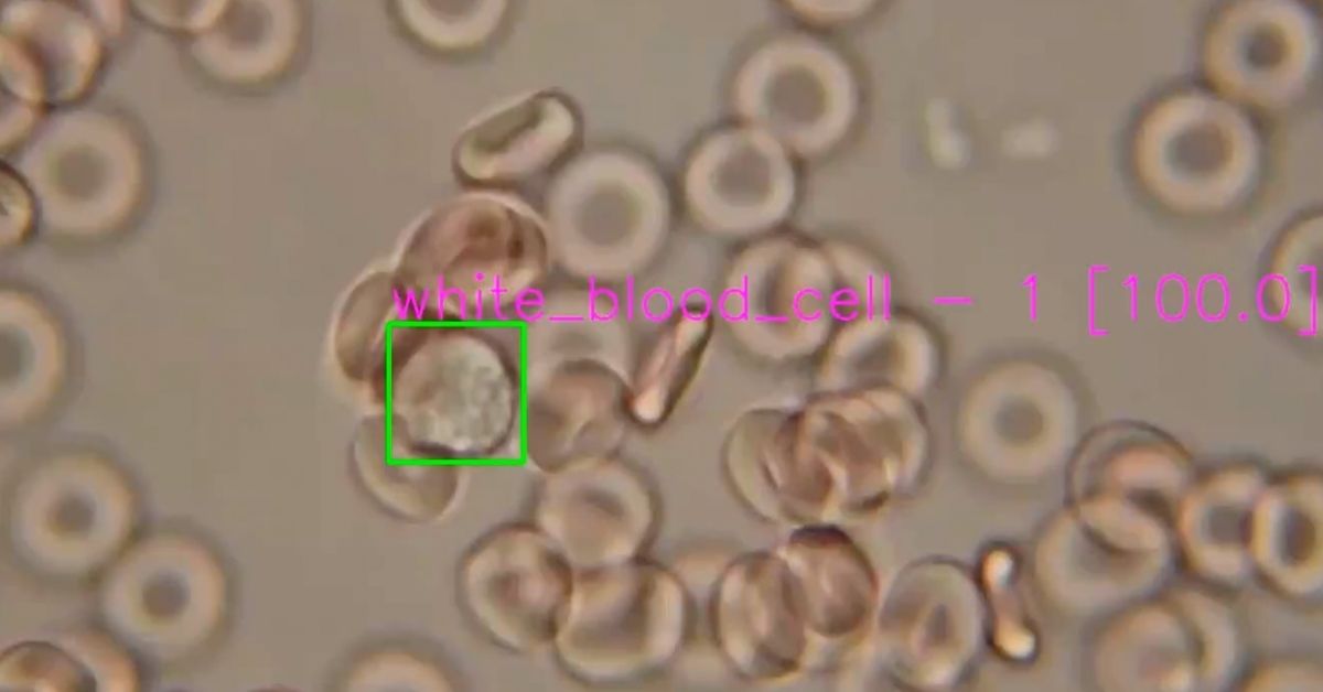
Computer vision technology is perfectly suited to automate the task of manually identifying and counting cells. For example, the Chooch AI platform is capable of identifying and counting cells with dramatically more accurate results than its human counterparts – even compared to the results of Ph.D. scientists and experienced research physicians.
The tremendous ROI benefits of computer vision technology for cell counting include:
- Faster cell counting results: Chooch AI completes 30-minute cell counting tasks in milliseconds, allowing the platform to achieve millions of human counting hours in just a few minutes.
- Improved accuracy: Chooch AI achieves 98% accuracy for most cell counting procedures. This is a striking improvement over the 80% standard for cell counting via traditional methods.
- Faster, better workflows: The speed, accuracy, and cost-efficiency benefits of computer vision for cell counting allow laboratories to analyze higher volumes of samples faster and more affordably.
- Labor cost savings: Chooch AI brings tremendous labor cost savings while freeing skilled scientists and doctors to devote their time to more important tasks.
- More competitive drug research: With pharmaceutical companies racing to test and release new drugs as quickly as possible, the efficiency of visual AI helps drug companies bring new medicines to market in record time.
- Better patient outcomes: Computer vision speeds the process of conducting a complete blood count analysis, and accurately counting blood, plasma, and lymph cells. This empowers healthcare practitioners to achieve better patient outcomes by reducing instances of delayed diagnoses and misdiagnoses.
- Lower sample dilution requirement: Computer vision solutions for cell counting are capable of accurately counting the cells in less diluted samples. By reducing the level of dilution, scientists can achieve more accurate counting results.
One of the primary advantages of Chooch AI is speed of implementation. With Chooch, laboratories can develop and deploy sophisticated computer vision models for cell counting in a matter of days. This is markedly faster than the six to nine months it generally takes to design and implement a visual AI model.
Chooch AI offers research laboratories immediate access to a wide library of pre-built computer vision models for the most common cell counting use cases. For more unique scenarios, laboratories can add layers of training to existing models – or train entirely new models from scratch – depending on the needs of the use case.
The ROI benefits of computer vision for cell counting are clear. With these new tools, medical labs diagnose patients faster and more accurately; drug companies develop new life-saving medications with greater efficiency; and, skilled scientists and doctors have extra time to devote to more pressing tasks.